Fluorescence imaging is one of the most widely used techniques in life science research. Its major strength lies in its ability to selectively and sensitively visualize specific cellular structures and biomolecules by labeling them with fluorescent probes and exciting them with light. However, fluorescence intensity is susceptible to various factors, such as fluctuations in excitation light intensity, local variations in fluorophore concentration, and photobleaching, which can compromise the accuracy and reliability of measurements. To address this challenge, fluorescence lifetime imaging, or fluorescence lifetime imaging microscopy (FLIM), has recently gained increasing attention. Fluorescence lifetime refers to the average time a fluorophore remains in its excited state before returning to the ground state. Unlike fluorescence intensity, it is relatively unaffected by such external factors, making it a more robust and quantitative indicator. However, lifetime measurements require time-resolved detection of fluorescence signals, which has traditionally limited the imaging speed of FLIM to video rates or much lower, making it difficult to capture fast cellular events or analyze large numbers of cells.
To overcome this limitation, we developed a technique for simultaneous multi-spot fluorescence lifetime measurement, achieving the world’s fastest FLIM at over 10,000 frames per second. In this approach, a continuous-wave laser beam is split into multiple beam spots, each modulated at a unique frequency and used to excite a distinct spatial point on the sample. While the emitted fluorescence signals are simultaneously detected by a single-pixel photodetector, the Fourier transformation allows for the separation and analysis of each signal due to its unique modulation frequency. The fluorescence lifetime at each spot is then estimated from the phase shift between the excitation and emission signals. Using this high-speed FLIM system, we conducted the world’s first large-scale fluorescence lifetime image analysis of heterogeneous cell populations, including tumors and cancer cells, demonstrating its broad utility. We are now actively exploring its applications in fields such as biomedicine and neuroscience, while also continuing to develop new fluorescence lifetime imaging technologies.
References
- H. Kanno, K. Hiramatsu, H. Mikami, A. Nakayashiki, S. Yamashita, A. Nagai, K. Okabe, F. Li, F. Yin, K. Tominaga, O. Bicer, R. Noma, B. Kiani, O. Efa, M. Büscher, T. Wazawa, M. Sonoshita, H. Shintaku, T. Nagai, S. Braun, J. P. Houston, S. Rashad, K. Niizuma, and K Goda, “High-throughput fluorescence lifetime imaging flow cytometry”, Nature Communications 15, 7376 (2024)
- H. Kanno, F. Li, J. Park, H. Endo, K. Niizuma, L. Gao, and K. Goda, "High-speed fluorescence lifetime imaging microscopy: techniques, applications, and prospects", Biophotonics Discovery 2, 030901 (2025)
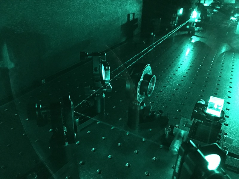
High-speed fluorescence lifetime imaging microscopy
- Field leader: Hiroshi Kanno
- Funding: JSPS KAKENHI, Takeda Science Foundation, Konica Minolta Foundation
- Collaboration: Serendipity Lab, Tohoku University Graduate School of Medicine